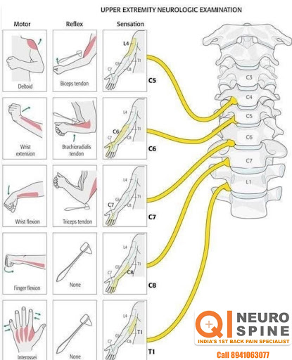
The is a diagram illustrating the upper extremity neurologic examination, which assesses the func…
The is a diagram illustrating the upper extremity neurologic examination, which assesses the function of the peripheral nervous system. It shows the relationship between specific spinal nerve roots and the motor, reflex, and sensation functions of the arm. Motor: The diagram links specific movements, such as wrist extension and flexion, to the corresponding nerve roots (e.g., C6 and C7). These movements are called myotomes, which are groups of muscles innervated by a single spinal nerve root. Reflex: It shows the reflexes tested in the upper arm, including the biceps, brachioradialis, and triceps reflexes, and their associated nerve roots (C5, C6, and C7, respectively). Sensation: The diagram illustrates the dermatomes, which are specific areas of the skin that are innervated by a single spinal nerve. This helps to assess sensory function, with the diagram showing the dermatomes for C5 through T1. Spinal Nerve Roots: The diagram shows the spinal cord and the cervical nerve roots (C3-T1) as they exit the vertebrae. Damage to these nerve roots can lead to a loss of a specific reflex or other neurological symptoms.